-
Before proceeding with
the actual placing,
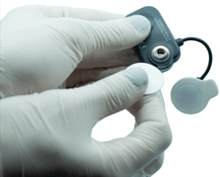
apply the pre-gelled electrodes using the provided snap connector and remove the protective film

-
Activate the probes
by swiping them on the
Swipe for activation
portion of the
charging station
Each probe is dedicated to the acquisition of a specific muscle, identified on the label:

Right Masseter:
![]()
Left Masseter:
![]()
Right Temporalis:
![]()
Left Temporalis:
![]()
Application of Probes to Temporalis muscles
To identify the anterior bundle of the temporalis, palpate the muscle by asking the patient to perform a full clench. Identify the major axis of the zygomatic process of the frontal bone and apply the probe along with the muscle anterior margin - close to the coronal suture and keeping 2 centimeters from the zygomatic process.

Application of Probes to Masseter muscles
To identify the masseter muscle, palpate the clenched muscle by identifying its belly. Apply the probe parallel to the course of the muscle fibers and in the central portion of the muscle - along the line joining the outside edge of the eye with the angle of the jaw.

Symmetry and Posture
Symmetry of placing between right and left should be maintained. To minimize any interference due to the patient's posture, ensure that the chair back is upright, and the patient is in a relaxed position with uncrossed legs, hands resting on the knees and looking straight forward.